Abstract
Achieving gross total resection is the primary prognostic factor in pilocytic astrocytomas. The spherical coordinate system (SCS) is an alternative to the Cartesian system for defining a point’s location in space. While the Cartesian system uses X, Y, and Z axes, the SCS utilizes ρ (distance from the origin), θ (the angle with the positive X-axis, 0 to 360 degrees), and ϕ (the angle with the positive Z-axis, 0 to 180 degrees). A 73-year-old female patient presented with headaches, dizziness, and gait disturbance over the past month, along with nausea and vomiting. She had previously undergone cerebellar tumor surgery in February 2020, and two years later, similar solid and cystic structures reappeared, suggesting a residual tumor. In such cases, using the SCS may help reduce remnant tumor rates and minimize the need for secondary operations.
Keywords: Cystic pilocytic astrocytoma, spherical coordinate system
INTRODUCTION
Thomas Stromer, quote “measure what can be measured, and make measurable what cannot be measured” attributed to Galileo; He simplifies it by saying “measure what needs to be measured” (). (In the same article, it is stated that this statement attributed to Galileo is not precise) Advanced mathematical calculations have been used in medicine for over a hundred years. Horsley and Clarke introduced the stereotaxy apparatus, which is a device that combined coordinate systems and anatomy, in 1908 (). Since many revolutionary advances such as stereotactic functional surgery, stereotactic radiosurgery, and associated biopsy have developed to the present day, these technologies have significantly improved patient outcomes. It has provided significant contributions to human life (-).
The dominant prognostic factor in pilocytic astrocytomas is the performance of gross total resection (). Mair et al. () showed that the localization of the lesion is critical for effective tumor resectability.
The spherical coordinate system (SCS) is one of the alternative methods used to define the location of a point in space (). The X, Y, Z directions are used in the Cartesian coordinate system. In the SCS, ρ, θ, and ϕ are defined as follows: ρ (radial distance) the from the origin , to be calculated, always greater than zero; θ (azimuth angle), the angle of ρ with the positive X-axis, which can take a value between 0 and 360 degrees; ϕ (elevation angle or polar angle), the angle that ρ makes with the positive Z-axis, which can take a value between 0 and 180 degrees 7. Our aim in presenting the case is to calculate the location of the preoperative solid mass with mathematical accuracy using a SCS. Our other aim is to present an alternative coordinate system to the Cartesian system used in stereotaxy.
CASE REPORT
A 73 year-old female patient with headache, dizziness, and gait disturbance in the last month. At the time of presentation, there was nausea and vomiting. On physical examination, she had dysmetria, dysdiadochokinesia and ataxia. The examination showed an arterial blood pressure of 120/75 mmHg, a pulse rate of 80/minute, a respiratory rate of 20 breaths/min, and an axillary temperature of 36.2 °C. She had a Glasgow Coma Scale (GCS) of 15. The patient was cooperative, with clear consciousness and oriented to time, place, and person. She did not have any weakness or neurological deficits. Also, coronary artery disease and diabetes mellitus were detected in anamnesis. The patient was comprehensively informed about the surgical procedure and its associated risks, and informed consent was obtained from the patient.
Surgical History
The patient had surgery for a cerebellar tumor in the second month of 2020 (, . The previous pathology report indicated that there was no tumor tissue. The same solid mass and cystic structures were observed after two years. In this case, firstly, the total impression of the remnant tumor was considered (, .
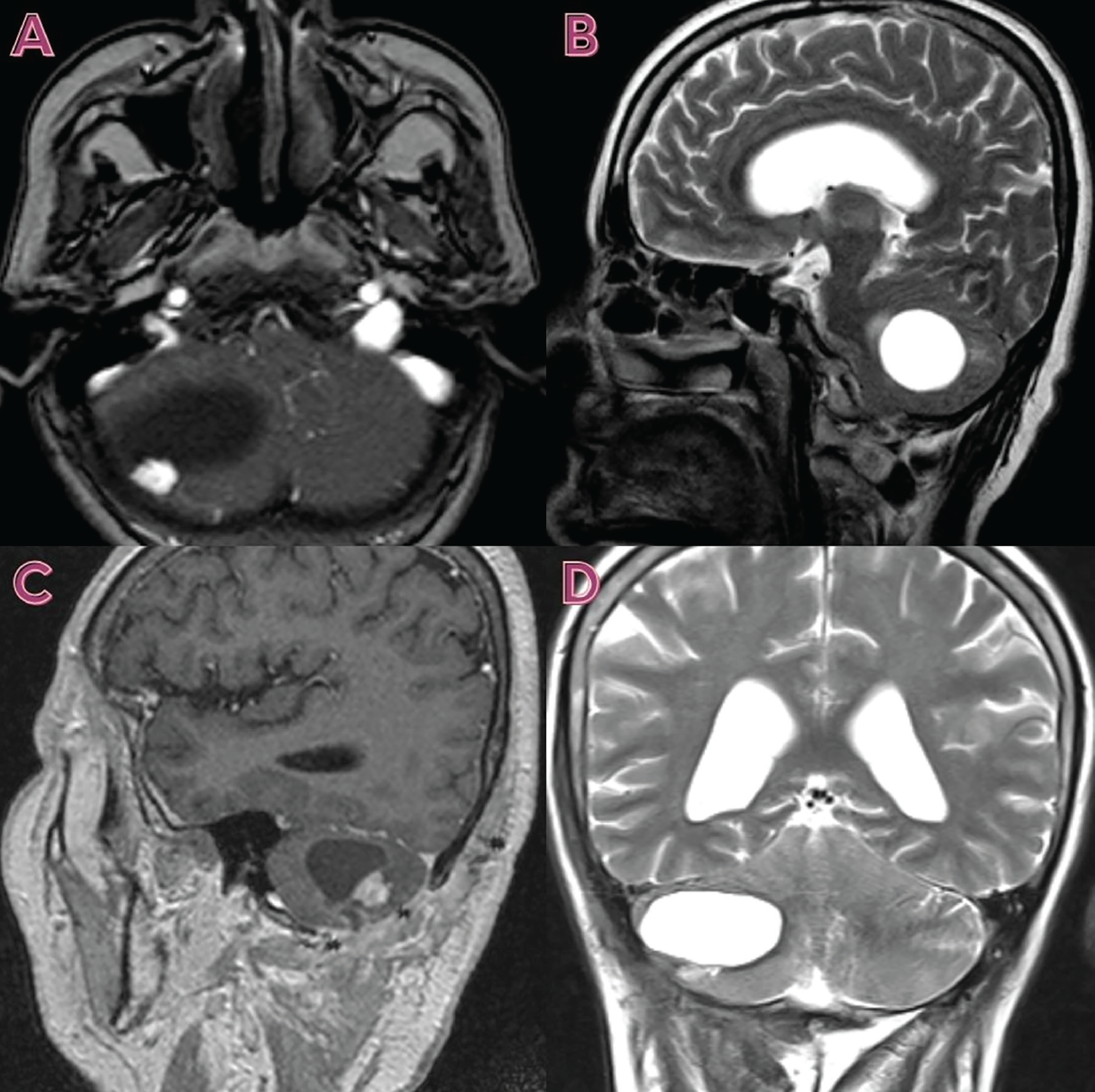
Preoperative Calculations with Imaging and Spherical Coordinate System
The patient’s cerebellar cystic space-occupying lesion resembles a sphere. The cerebellar cystic lesion was considered to be spherical, and an attempt was made to reach the solid lesion. With the help of the SCS, the coordinates of the solid lesion were calculated according to the point designated as the center of the sphere. The distance of the solid lesion to the center of the cyst; That is, the “ρ” value, was approximately 12 mm (). The angle of the solid lesion with the x-axis of the cyst, that is, the θ value, was calculated between 300° and 346° (). The angle of the solid lesion with the z-axis of the cyst, that is, the ϕ value was calculated between 97° and 144° (). The coordinates of the patient’s solid lesion were calculated as r, θ1- θ2, ϕ1- ϕ2 values according to the accepted center of the cyst: 12 mm, 300°-346°, 97°-144°.
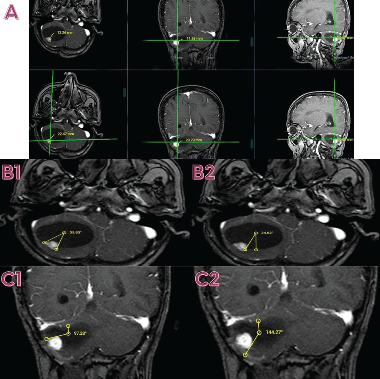
Surgical Technique
The surgery was started with precise calculations in the pre-operative stage. The tumor was completely resected in approximately 70 minutes. The patient was taken to the intensive care unit in the early post-operative stage. However, the patient’s GCS decreased to 12 after 2 hours. Control brain tomography shows cerebellar edema and mild brainstem compression. The patient was started on 600 mg mannitol. After medical treatment, the patient’s GCS rose again to 15 on the postoperative second day. Mannitol treatment was terminated after gradually reducing the dose. The patient was taken to the service on the 5th postoperative day. On the 15th postoperative day, the patient was discharged with full recovery. The reason the patient is monitored for such a long time is the imbalance in blood pressure and glucose levels. In this process, necessary branch consultations were conducted.
Postoperative control brain magnetic resonance imaging (MRI) examinations of the patient showed that the total tumor resection was successful and the cyst had disappeared (). Pilocytic astrocytoma, WHO grade 1 was detected in histopathological examination ().
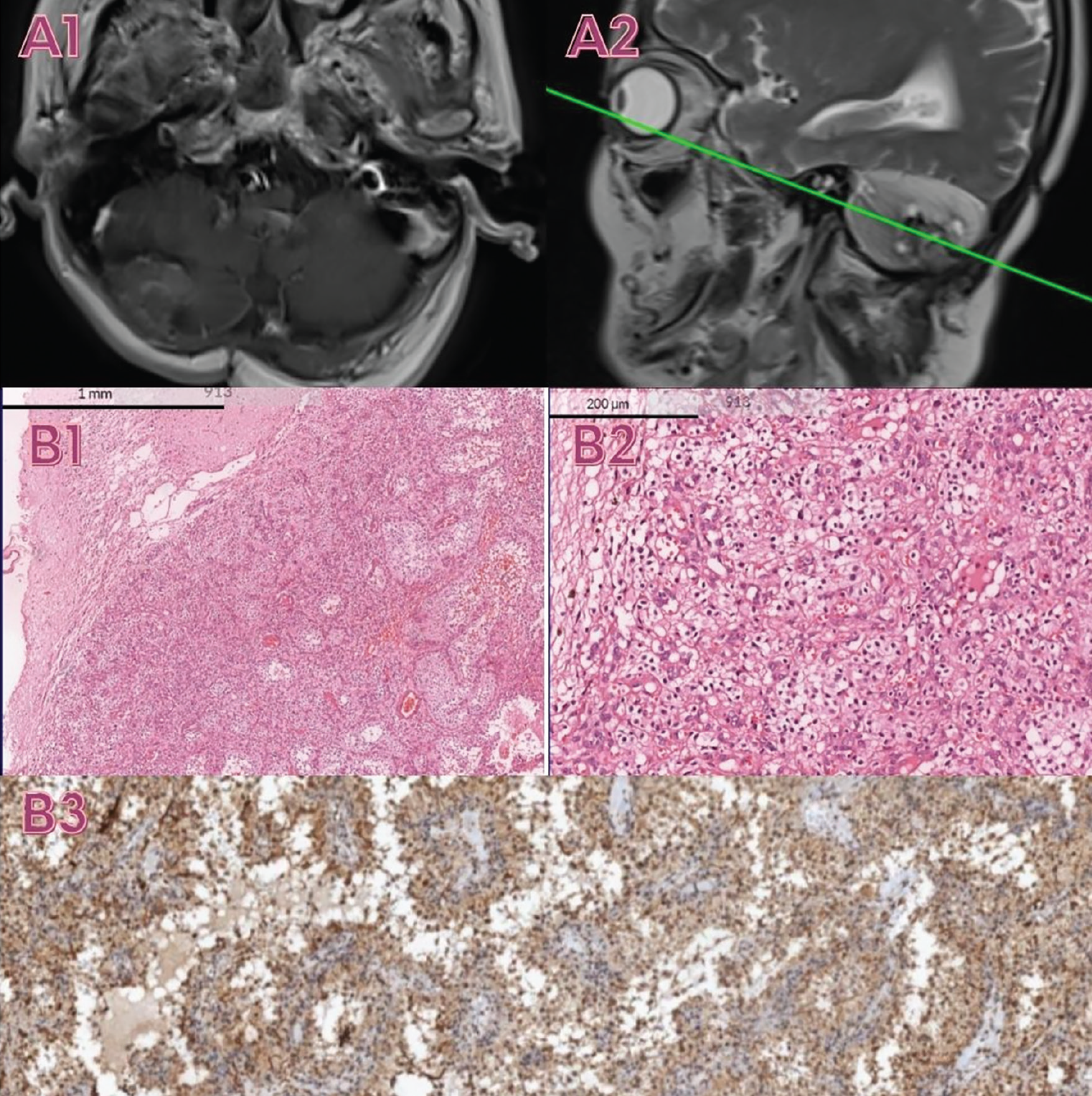
GFAP; Glial fibrillary acidic protein
A neoplastic proliferation with a rich capillary vascular network within the cerebellum was remarkable in hematoxylin-eosin sections. This lesion had no distinctive capsule structure but was well demarcated. Neoplastic cells were clear, polygonal or rounded, with abundant cytoplasm and revealed no prominent nucleoli ( B1,B2). These neoplastic cells were positive for glial fibrillary acidic protein ( B3), S100, and ATRX and negative for OLIG-2 and Neu-N. The proliferation index was about 1-2% as determined with the marker Ki67 (not shown). When clinical, radiological, and histopathological findings were evaluated together, the case was diagnosed as pilocytic astrocytoma/WHO grade 1.
DISCUSSION
As far as we could detect, the finding of tumor localization using the SCS was reported in the literature for the first time. The SCS can also be used as an alternative in case management, just like the Cartesian coordinate system. This may reduce the rates of remnant tumors and secondary operations. This case report provides a foundation for a more in-depth study of SCS and its clinical outcomes.
However, a study in the engineering literature shows Cheng joint rotations with an SCS ().
Techniques such as intraoperative MRI are available to guide surgeons during surgery (). Several techniques are beneficial during surgery, depending on the pathology of the tumor, such as intraoperative ultrasound navigation, 5-aminolevulinic acid, and Fluorescein Na, and intraoperative computed tomography (). The mathematical calculations that we used to find the tumor’s address can potentially be used alone or in combination with the techniques we mentioned above ().
After reaching the cyst, we were able to immediately locate the solid tumor, whose spherical coordinates we calculated. We completely removed the solid lesion by circumferential excision. It can be simplified in an illustration to make the mathematical calculations easier (). A cystic sphere can be divided into 8 chambers. The locations of these chambers can be evaluated with MRI. Sphere-like cyst: 8 chambers as superior-lateral-posterior, superior-lateral-anterior, inferior-lateral-posterior, inferior-lateral-anterior, superior-medial-posterior, superior-medial-anterior, inferior-medial-posterior, inferior-medial-anterior are separable. Solid mass was calculated in the inferior, lateral, and posterior chambers of the cyst (, ). Cystic cerebellar hemangioblastomas that may have similar radiological characteristics due to remnant tumors have been reported in literature (). The mathematical system we used could be useful in locating tumors. It can reduce the amount of residual and remnant tissue.
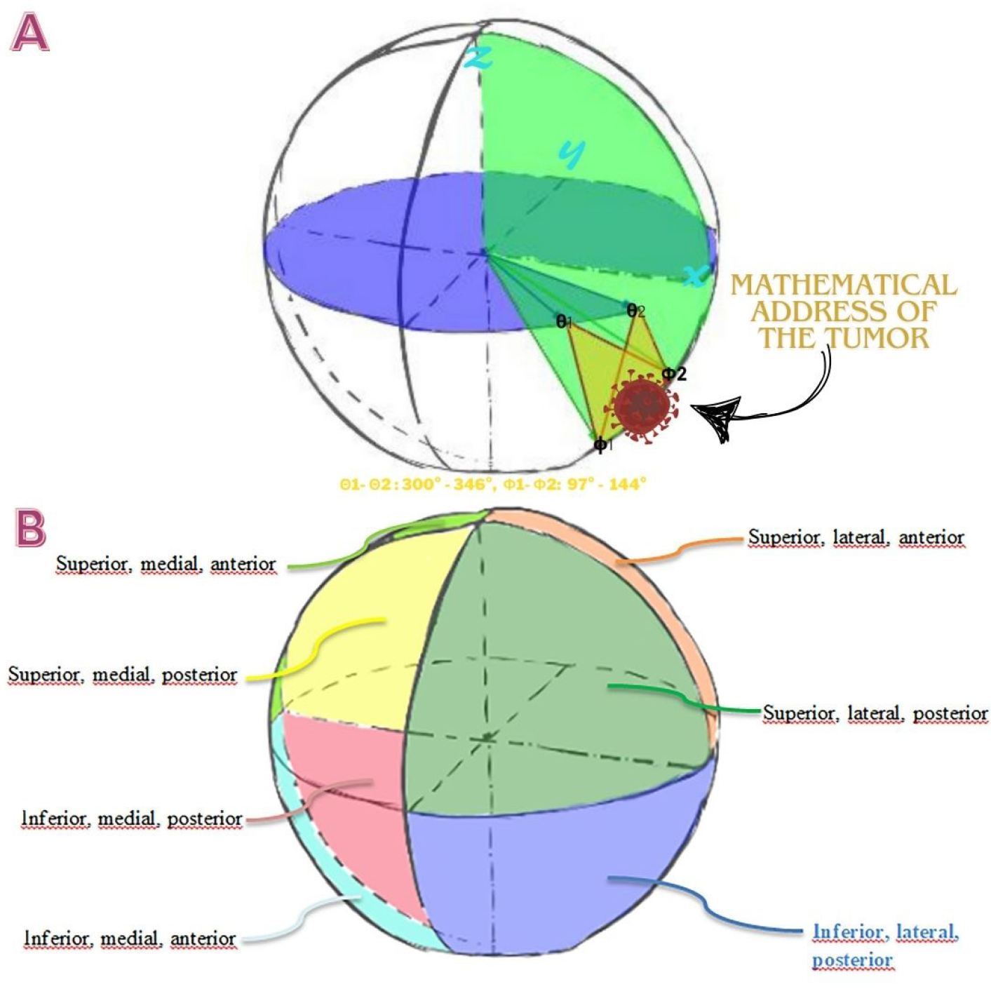
Study Limitations
Our study has some limitations, and it progresses by initially accepting the cyst as a sphere. We are aware that the cyst is not a complete sphere. Since the cyst was not a complete sphere, the coordinates of the solid mass were calculated approximately. However, even this calculation was sufficient to locate the tumor. The presented study is based on a single case, limiting the generalizability of findings. A broader patient cohort would be necessary to validate the consistency and efficacy of the SCS approach across different anatomical variations.
CONCLUSION
The article lacks a comparative analysis with traditional stereotactic methods. A comparative study would provide a clearer understanding of the advantages and disadvantages of the SCS in contrast to existing techniques.
Footnotes
Ethics
Informed Consent: The patient was comprehensively informed about the surgical procedure and its associated risks, and informed consent was obtained from the patient.
Authorship Contributions
Surgical and Medical Practices: A.E.T., Ö.E.S., Concept: A.E.T., Design: A.E.T., Data Collection or Processing: R.B.G., Analysis or Interpretation: A.E.T., Ö.E.S., Literature Search: A.E.T., Writing: A.E.T., R.B.G.
Conflict of Interest: No conflict of interest was declared by the authors.
Financial Disclosure: The authors declare that this study received no financial support.
References
T. Strohmer. “Measure what should be measured: progress and challenges in compressive sensing,” in IEEE Signal Processing Letters, 2012;19:887-93.
Yang I, Udawatta M, Prashant GN, Lagman C, Bloch O, Jensen R, et al. Stereotactic radiosurgery for neurosurgical patients: a historical review and current perspectives. World Neurosurg. 2019;122:522-31.
Ostertag CB, Mennel HD, Kiessling M. Stereotactic biopsy of brain tumors. Surg Neurol. 1980;14:275-83.
Spiegel EA, Wycis HT, Marks M, Lee AJ. Stereotaxic apparatus for operations on the human brain. Science. 1947;106:349-50.
Villanueva KG, Rea ND, Krieger MD. Novel surgical and radiologic risk factors for progression or recurrence of pediatric pilocytic astrocytoma. Pediatr Neurosurg. 2019;54:375-85
Mair MJ, Wöhrer A, Furtner J, Simonovska A, Kiesel B, Oberndorfer S, Ungersböck K, Marosi C, Sahm F, Hainfellner JA, Rössler K, Preusser M, Widhalm G, Berghoff AS. Clinical characteristics and prognostic factors of adult patients with pilocytic astrocytoma. J Neurooncol. 2020;148:187-98.
Reed BC, editor. Keplerian Ellipses: The Physics of the Gravitational Two-body Problem. 1st ed. Morgan & Claypool Publishers; 2019.
Cheng PL. A spherical rotation coordinate system for the description of three-dimensional joint rotations. Ann Biomed Eng. 2000;28:1381-92.
Choudhri AF, Siddiqui A, Klimo P Jr, Boop FA. Intraoperative MRI in pediatric brain tumors. Pediatr Radiol. 2015;45 Suppl 3:S397-405.
Tejada Solís S, de Quintana Schmidt C, Gonzalez Sánchez J, Fernández Portales I, Del Álamo de Pedro M, Rodríguez Berrocal V, et al. Intraoperative imaging in the neurosurgery operating theatre: a review of the most commonly used techniques for brain tumour surgery. Neurocirugia (Astur : Engl Ed). 2020;31:184-94.
Cervio A, Villalonga JF, Mormandi R, Alcorta SC, Sevlever G, Salvat J. Surgical treatment of cerebellar hemangioblastomas. Surg Neurol Int. 2017;8:163.