Abstract
Ovarian tumors have a wide spectrum and are pathologically classified in four main groups as epithelial, germ cell, sex cord-stromal, and metastatic tumors. Our aim is to demonstrate magnetic resonance imaging findings of benign ovarian masses with illustrative cases.
Epithelial Tumors
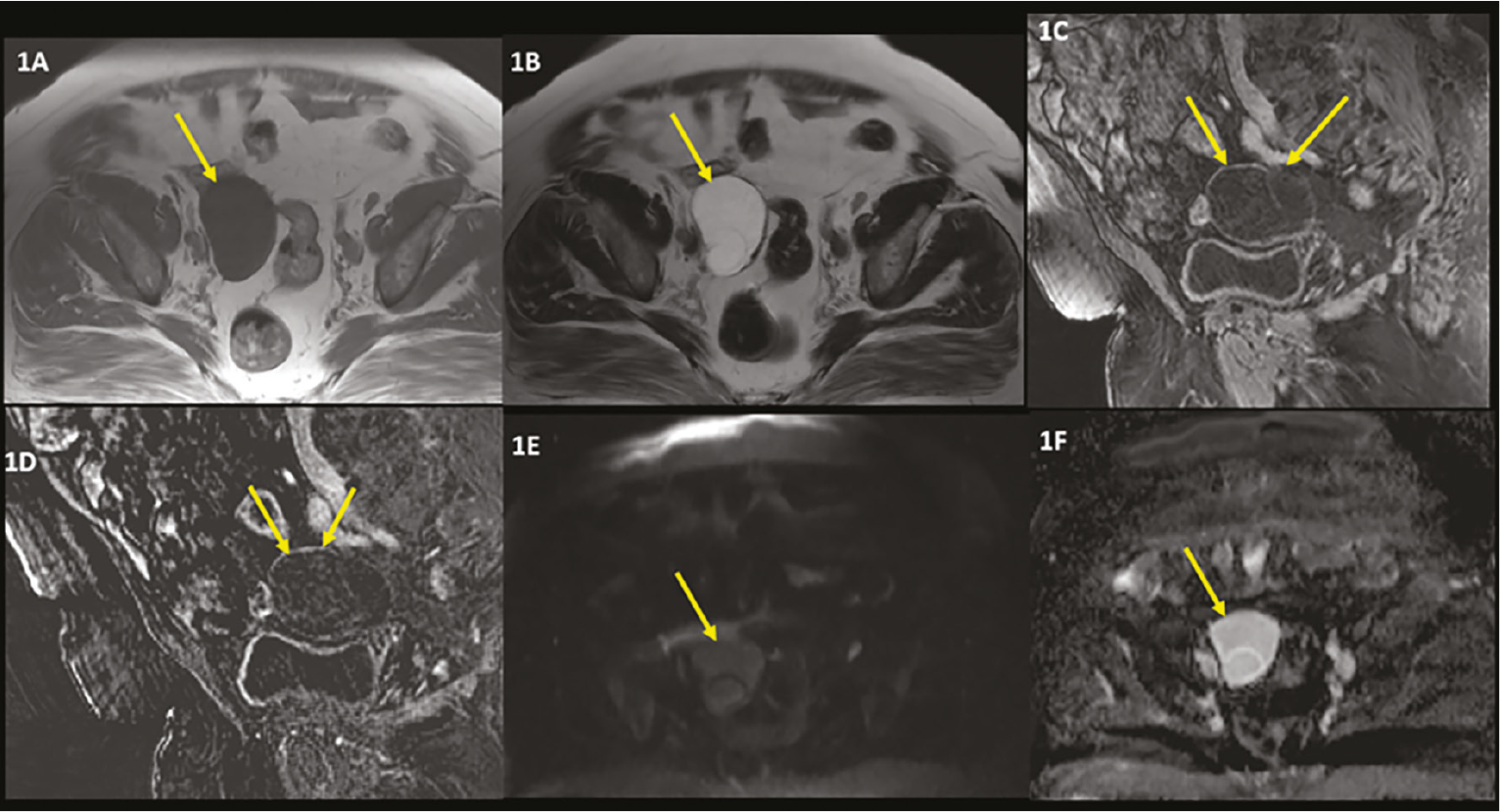
• Serous cystadenoma is the most common epithelial tumor. Case 1 is serous cystadenoma.
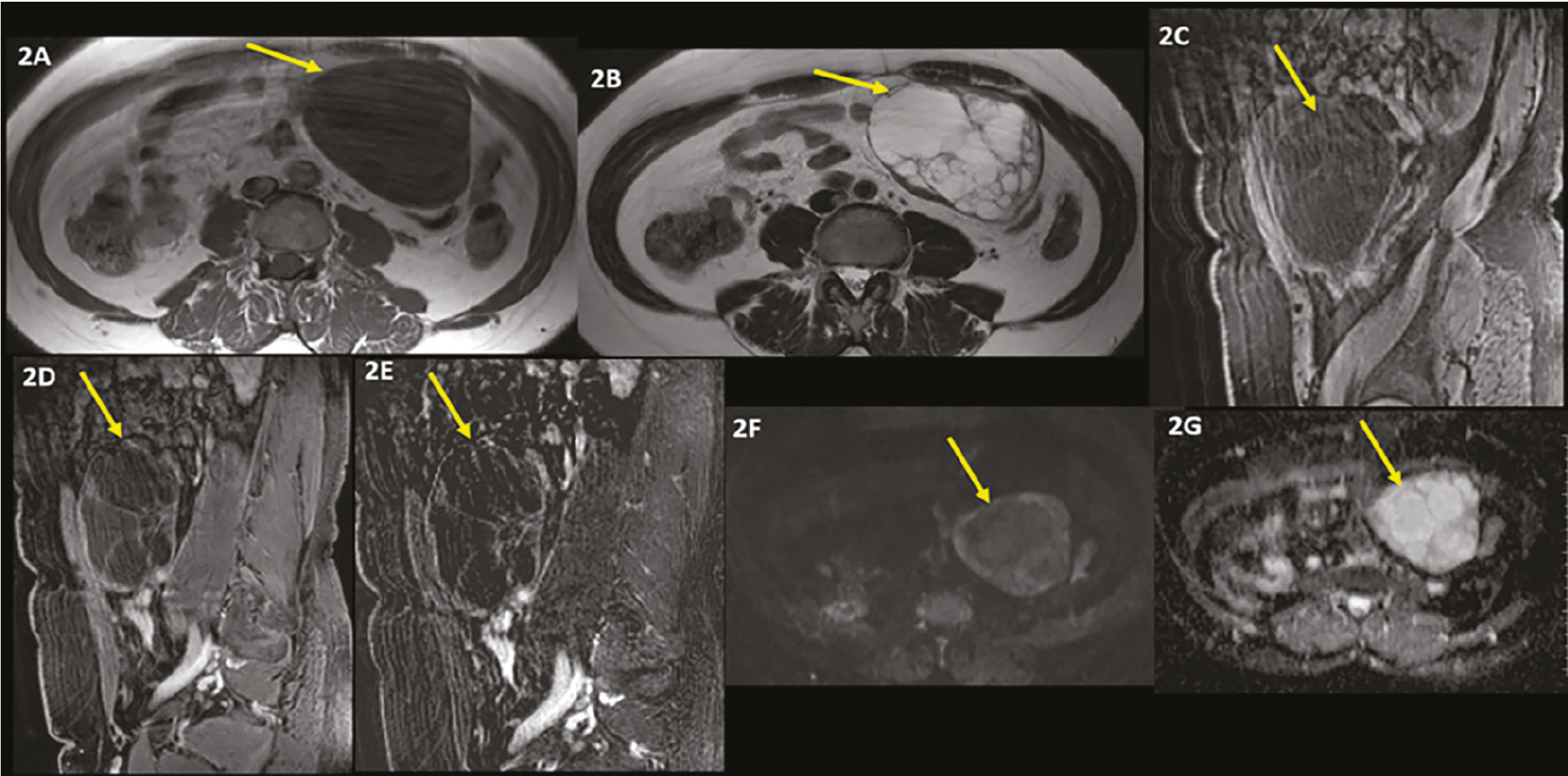
• Mucinous cystadenomas are often unilateral and tend to be larger cystic lesions. Case 2 is a mucinous cystadenoma.
Germ Cell Tumors
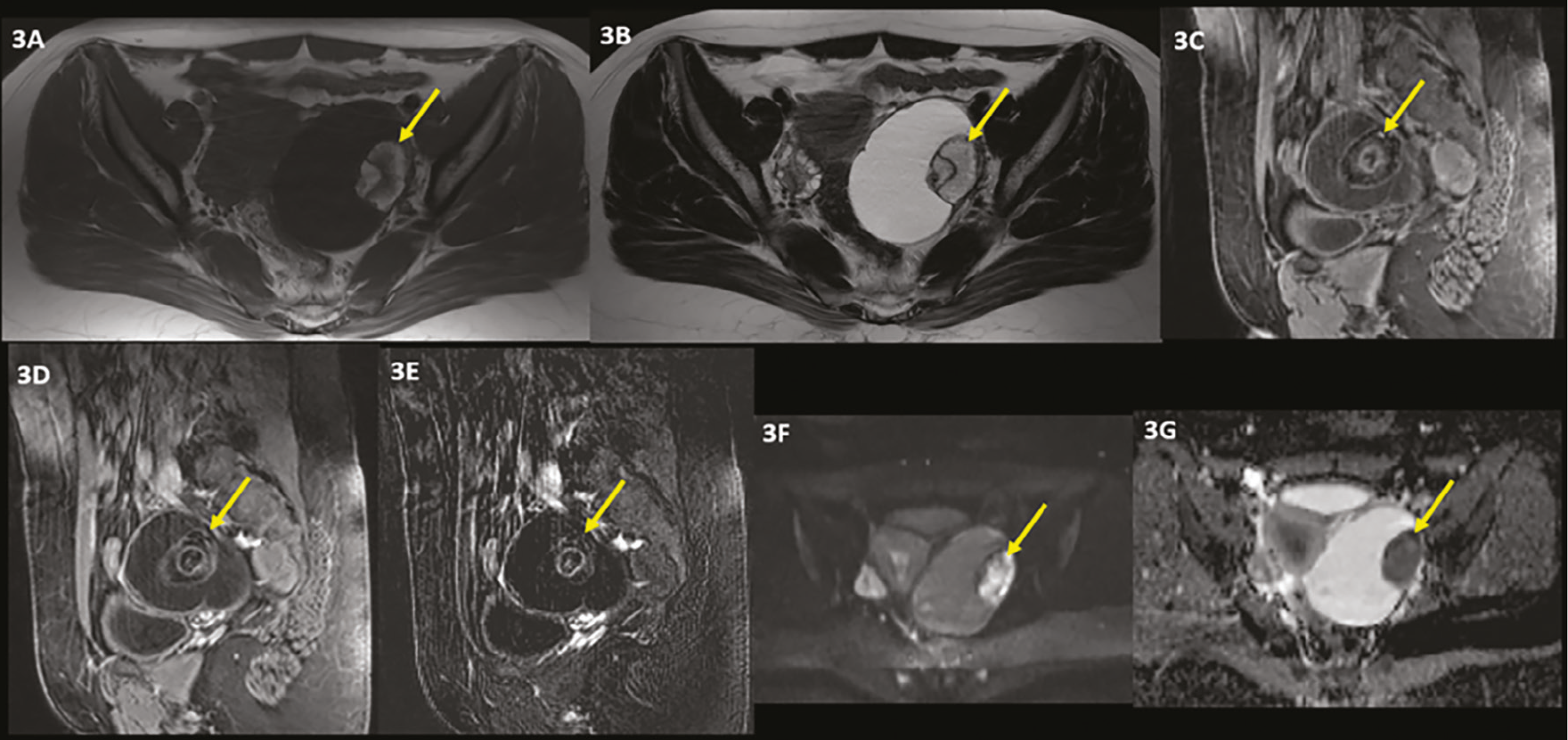
• Mature teratomas are unilocular masses filled with oily material and their walls contain hair follicles, skin glands, etc. Case 3 is mature teratoma.
Sex Cord-Stromal Tumors
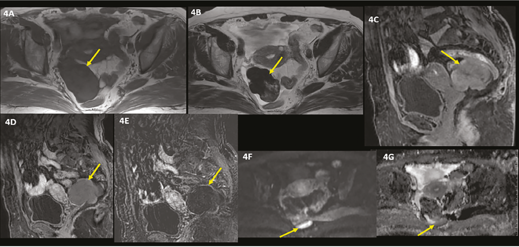
• Fibrothecoma is the most common sex cord tumor. Case 4 is fibrothecoma.
• Cystadeofibroma is an uncommon benign tumor in which fibrous stroma is the dominant component. Case 5 is cystadenofibroma.
• Sclerosing stromal tumor is a rare benign tumor. Case 6 is a sclerosing stromal tumor.
Keywords: Adnexial benign lesions, magnetic resonance imaging, epithelial tumors
INTRODUCTION
Ovarian tumors have a wide spectrum and are pathologically classified into four main groups: epithelial, germ cell, sex cord-stromal tumors, and metastatic tumors ().
The most common group is epithelial cancer. Epithelial benign cancers include serous cystadenoma, mucinous cystadenoma, cystadenofibroma, and Brenner tumors. Benign germ cell tumors include mature cystic teratomas. Benign sex cord-stromal tumors include thecoma, fibroma, fibrothecoma, and sclerosing stromal tumor ().
The goal of our presentation is to demonstrate magnetic resonance imaging (MRI) findings of benign ovarian masses with illustrative cases.
CASE REPORT
It was stated that the names of the cases presented in case reports would not appear, and that permission was obtained for publication. Written and verbal consent was obtained from all patients/legal representatives.
Patients whose ultrasonographic (US) examinations revealed adnexal masses were referred to our department for lesion characterization with MRI.
Our MRI protocole consisted of big field of view (FOV) axial and coronal T1 weighted turbo spin-echo, small FOV and high resolution T2-weighted (T2W) turbo spin-echo sequences in 3 different planes, axial T2W echoplanar imaging, sagittal pre- and dynamic post-contrast gradient-echo T1-weighted (T1W) sequence and subtraction images (, ).
Epithelial Tumors
• Serous Cystadenoma: It is the most common epithelial ovarian tumor, and 15-20% of serous cystadenomas are bilateral. In MRI, it has low-medium intensity on T1W and high-intensity on T2W images as a pure cyst and sometimes has small millimetric papillary projections. Often, thin <3 mm septa can be seen (-). Their average diameter is 10 cm, while 30 cm in diameter cystadenomas are reported in the literature. Case 1 was pathologically proven to be a serous cystadenoma ().
• Mucinous Cystadenoma: They are often unilateral, multiloculated, and tend to be larger cystic lesions. They often contain only thin walls and septa. Depending on the density of mucinous content, lesions’ T1W and T2W signals may differ. The presence of a thick septum (>5 mm) or a wall or a solid component with contrast enhancement should raise the suspicion of malignancy ().
Case 2 was pathologically proven to be a mucinous cystadenoma ().
Germ Cell Tumors
• Mature Teratoma: Mature cystic teratomas are unilocular masses filled with oily material and their walls contain hair follicles, skin glands, muscles, etc. Imaging findings, may range from a pure cyst to a mixed mass containing components of 3 germ sheets or a solid non-cystic mass containing mostly fat.
In MRI, hyperintensity T1W and suppression hyperintensity in fat-suppressed T1W sequence and variable fat-induced hyperintensity in the T2W sequence is typical ().
Fat-suppressed T1W sequence is the key point in MRI diagnosis (). Teeth and calcifications can be recognized as hypointense in all sequences. Ovarian teratomas can be associated with various complications including torsion (16% of ovarian teratomas), rupture (1-4%), malignant transformation (1-2%), infection (1%). Due to rupture, granulomatous peritonitis may occur.
Case 3 was pathologically proven to be a mature teratoma ().
Sex Cord-Stromal Tumors
• Fibrothecoma: It is the most common sex cord tumor seen in the pre-post menopausal period. On MRI, the low signal in T1W and very low signal in T2W sequences are typical diagnostic features (). Scattered high-signal areas within the mass may be observed due to edema or degeneration. Case 4 was pathologically proven to be a fibrothecoma ().
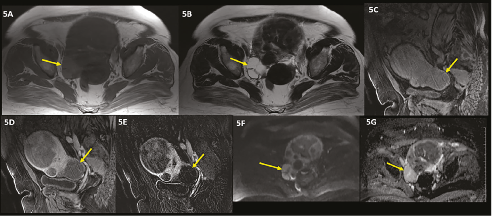
• Cystadeofibroma: Cystadeofibroma is an uncommon benign tumor in which fibrous stroma is the dominant component of the lesion. They have either a pure cystic or complex cystic pattern with nodular or trabecular pattern ().
On MRI, in T2W sequences fibrous stromal components such as septa have low signal intensity while cystic components have high signal and this composition creates the “black sponge” like appearance. Septa may show moderate contrast enhancement.
Case 5 was pathologically proven to be a cystadenofibroma ().
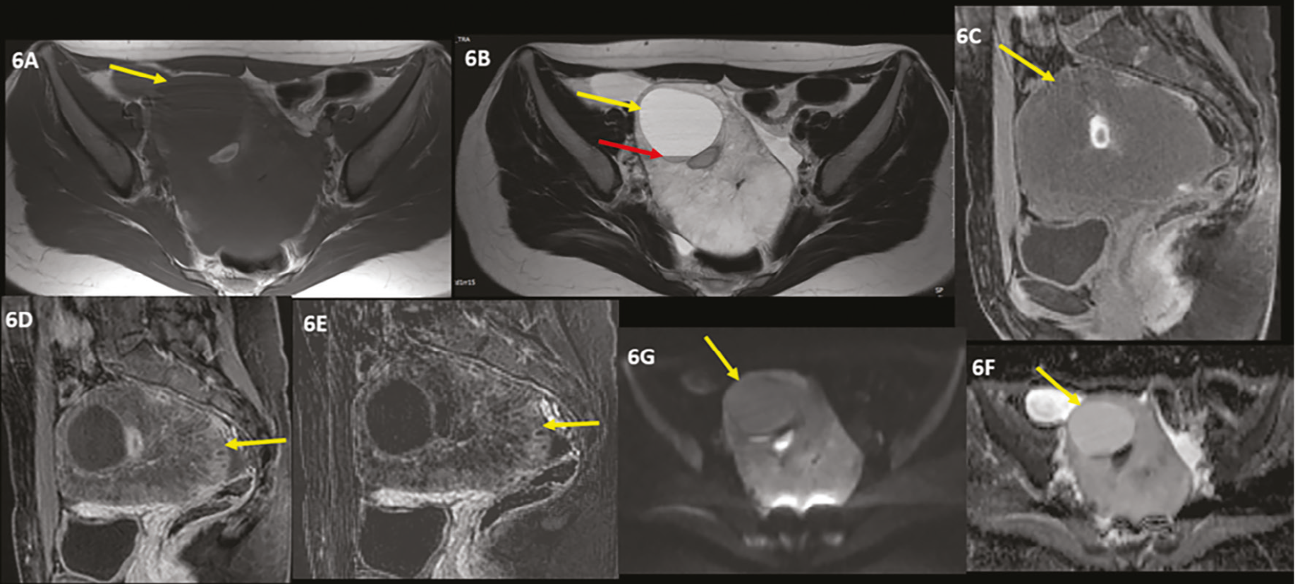
• Sclerosing Stromal Tumor: Sclerosing stromal tumor is a rare benign ovarian tumor that occurs in young women in the second and third decades. On MRI, on T2W sequences, it is a large mass with a solid heterogeneous component containing hyperintense cystic areas and a medium-high signal (). Contrast-enhanced MRI shows early peripheral and progressive centripetal enhancement
Case 6 was pathologically proven to be a sclerosing stromal tumor ().
DISCUSSION
MRI is regarded as a problem-solving method for evaluating adnexal masses. When evaluating adnexal masses, gadolinium-enhanced MRI tends to be more accurate than US.
Even if solid components (such as Rokitansky nodules) are also present, the presence of fat in a cystic adnexal lesion is indicative of a cystic teratoma. Both conventional and fat-suppressed T1W imaging are necessary to demonstrate fat since the latter helps distinguish fat from blood products as the source of the high T1 signal intensity (). Nonetheless, ovarian fibromas may be better characterized by T2W imaging. To evaluate a clinically suspected adnexal mass, US is still the predominant imaging modality, according to established practice and a review of the literature. If the outcome of the US evaluation is uncertain, MRI represents a financially advantageous course of action. Complex adnexal masses can be detected and characterized with great detection and characterization capabilities using gadolinium-enhanced MRI, and it also exhibits strong inter- and intraobserver agreement.
In case of a suspected adnexal mass, pelvic or transvaginal US is the first-step imaging modality. However, MRI’s high potential for tissue characterization makes it the second step and a decisive imaging modality. MRI diagnosis of benign ovarian masses is critical in the management of patients affected by them.
Footnotes
Ethics
Informed Consent: Written and verbal consent was obtained from all patients/legal representatives.
Acknowledgment
This article was previously published as a poster at the European Congress of Radiology 2023.
Authorship Contributions
Surgical and Medical Practices: A.K.İ., S.G., Concept: A.K.İ., S.G., Design: A.K.İ., S.G., B.A., Data Collection or Processing: A.K.İ., S.G., B.A., Analysis or Interpretation: A.K.İ., Literature Search: A.K.İ., Writing: A.K.İ., S.G.
Conflict of Interest: No conflict of interest was declared by the authors.
Financial Disclosure: The authors declare that this study received no financial support.
References
Pekindil G. Over tümörlerinde görüntüleme. Trd Sem. 2015;3:108-26.
Jeong YY, Outwater EK, Kang HK. Imaging evaluation of ovarian masses. Radiographics. 2000;20:1445-70.
Kurtz AB, Tsimikas JV, Tempany CM, Hamper UM, Arger PH, Bree RL, et al. Diagnosis and staging of ovarian cancer: comparative values of doppler and conventional US, CT, and MR imaging correlated with surgery and histopathologic analysis report of the Radiology Diagnostic Oncology Group. Radiology. 1999;212:19-27.
Siegelmann ES, Outwater EK. Tissue characterization in the female pelvis by means of MR imaging. Radiology. 1999;212:5-18.
Imaoka I, Wada A, Kaji Y, Hayashi T, Hayashi M, Matsuo M, et al. Developing an MR imaging strategy for diagnosis of ovarian masses. Radiographics. 2006;26:1431-48.
Hricak H, Chen M, Coakley FV, Kinkel K, Yu KK, Sica G, et al. Complex adnexal masses: detection and characterization with MR imaging. Radiology. 2000;214:39-46.
Thomassin-Naggara I, Aubert E, Rockall A, Jalaguier-Coudray A, Rouzier R, Daraï E, et al. Adnexal masses: development and preliminary validation of an MR imaging scoring system. Radiology. 2013;267:432-43.
Atram M, Anshu, Sharma S, Gangane N. Sclerosing stromal tumor of the ovary. Obstet Gynecol Sci. 2014;57:405-8.